Renal Artery Ultrasound
What Is a Renal Artery Ultrasound?
How to Prepare for a Renal Artery Ultrasound?
Typically, no special preparation is required for a renal artery ultrasound, but your healthcare provider may give specific instructions based on your individual needs. Here are some general guidelines:
- Wear comfortable, loose-fitting clothing.
- Avoid clothing with metal zippers, snaps, or buttons in the area being examined.
- You may be instructed to fast for a certain period before the exam to prevent gas buildup in the intestines. Your provider will inform you if fasting is necessary and for how long.
- If you're taking any medications, including blood pressure medications, your provider may ask you to temporarily stop taking them before the exam.
- Inform your healthcare provider about any allergies to latex, ultrasound gel, or other substances.
- You may be asked to drink several glasses of water before the exam to fill your bladder, which can enhance the quality of the images.
Why Is a Renal Artery Ultrasound Performed?
A Renal Artery Ultrasound, also called a renal artery duplex, is commonly performed in individuals with hypertension to evaluate the renal arteries. These arteries supply blood to the kidneys, and if they become narrowed due to stenosis or plaque buildup, it can result in high blood pressure. The ultrasound helps screen the condition of the arteries, identify any blockages or plaque accumulation that may cause narrowing, and assess for potential aneurysms in the abdominal aorta.
What to Expect During a Renal Artery Ultrasound?
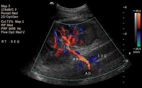
The renal artery duplex scan uses an ultrasound probe to examine your kidneys and the arteries that supply blood to them. During the procedure, the technician will view the images on a computer screen. As the transducer is placed on your abdomen, you may feel some pressure, but the exam is painless and non-invasive. The procedure usually takes about 10 minutes to complete.
Follow-Up After a Renal Artery Ultrasound
Following the renal artery ultrasound, healthcare providers will review the images to assess the condition of the renal arteries, looking for any plaque or blockages, and evaluating the vessels’ integrity. Based on the results, further 3D imaging may be recommended. The exam might also uncover incidental findings, such as kidney cysts or stones, which may require further evaluation with a dedicated renal/kidney ultrasound. If necessary, you’ll be advised to follow up with a nephrologist or urologist.
Potential Risks of a Renal Artery Ultrasound
There are no known risks associated with renal artery ultrasound. It is a non-invasive, safe, and painless procedure that uses sound waves to create images of the renal arteries and kidneys. Unlike imaging tests that use radiation or contrast agents, this ultrasound does not involve exposure to harmful radiation or substances. It is considered a low-risk procedure, and any discomfort or pressure felt during the exam is temporary and typically subsides after the procedure.
Are There Any Related Tests to a Renal Artery Ultrasound?
Yes, there are several tests related to a renal artery ultrasound, including:
- Renal MRI or CT Scan: These imaging tests offer more detailed views of the renal arteries and can help identify other abnormalities, such as tumors or cysts.
- Renal Nuclear Medicine Scan: This test uses a small amount of radioactive material to assess kidney function and blood flow.
- Renal Angiogram: This procedure involves injecting contrast dye into the renal arteries and using X-ray imaging to evaluate blood flow and detect blockages or narrowing. It is more invasive than a renal artery ultrasound and carries a higher risk of complications.